
Neuropoli
Modern Medicine. Compassionate Care.
Book Appointment
Neuropoli
Modern Medicine. Compassionate Care.
Book Appointment
Neuropoli
Modern Medicine. Compassionate Care.
Book Appointment
Our Diagnostics
Modern and Specialized Neurological Diagnostics
Here you will find an overview of our diagnostic procedures and specialized examinations.
Ultrasound of the Neck and Brain Arteries
Neurovascular ultrasound is a safe, painless and radiation-free method for examining the arteries supplying the brain. Using Doppler and duplex ultrasound, we can assess the arteries of the neck and brain and detect vascular changes and blood flow abnormalities. The examination therefore plays an important role in stroke diagnosis and prevention.
We use state-of-the-art ultrasound systems, including the Philips EPIQ Elite and the Viasonix Dolphin 4D with a robot-assisted XF probe for examining the cerebral arteries through the skull (transcranial Doppler ultrasound). In addition, we offer specialized neurovascular ultrasound techniques.
Specialized Neurovascular Ultrasound
Specialized neurovascular ultrasound complements the conventional examination of the arteries supplying the brain. Using the robot-assisted XF probe of the Viasonix Dolphin 4D, we can assess cerebral blood flow and its regulation in greater detail and detect right-to-left shunts and microemboli.
Assessment of cerebrovascular reserve (CVR) evaluates how effectively the cerebral blood vessels can increase blood flow when needed. It provides important information in patients with narrowing or occlusion of the arteries supplying the brain and in moyamoya disease and moyamoya syndrome. In selected patients, it can also help assess cerebral hemodynamics when considering revascularization procedures such as EC-IC bypass surgery.
Assessment of cerebral autoregulation evaluates the brain’s ability to maintain relatively stable blood flow despite fluctuations in blood pressure.
PFO assessment using a bubble test (contrast-enhanced ultrasound) is used to detect a right-to-left shunt, particularly a patent foramen ovale (PFO). It plays an important role in the evaluation of strokes without an identifiable cause as well as in specific diving medicine assessments.
Microemboli detection uses transcranial Doppler ultrasound to detect tiny circulating emboli over an extended period and can help identify potential sources of embolism.
Electrophysiological Diagnostics
Electrophysiological diagnostics assess the function of nerves, muscles, and central nervous pathways. They help detect, localize, and evaluate a wide range of neurological disorders.
The main procedures include nerve conduction studies (NCS) to assess nerve function, electromyography (EMG) to evaluate muscle function, and evoked potentials (EP) to assess the visual pathways (VEP), auditory pathways (AEP), and sensory pathways (SEP).
We also assess the autonomic nervous system, which regulates functions such as circulation, heart rate, and sweating. Autonomic dysfunction may occur, for example, in polyneuropathy and diabetes mellitus, in patients with orthostatic symptoms and POTS, and in association with ME/CFS. For autonomic function testing, we use the sympathetic skin response (SSR) and heart rate variability (HRV) analysis to assess the autonomic regulation of heart rate and circulation.
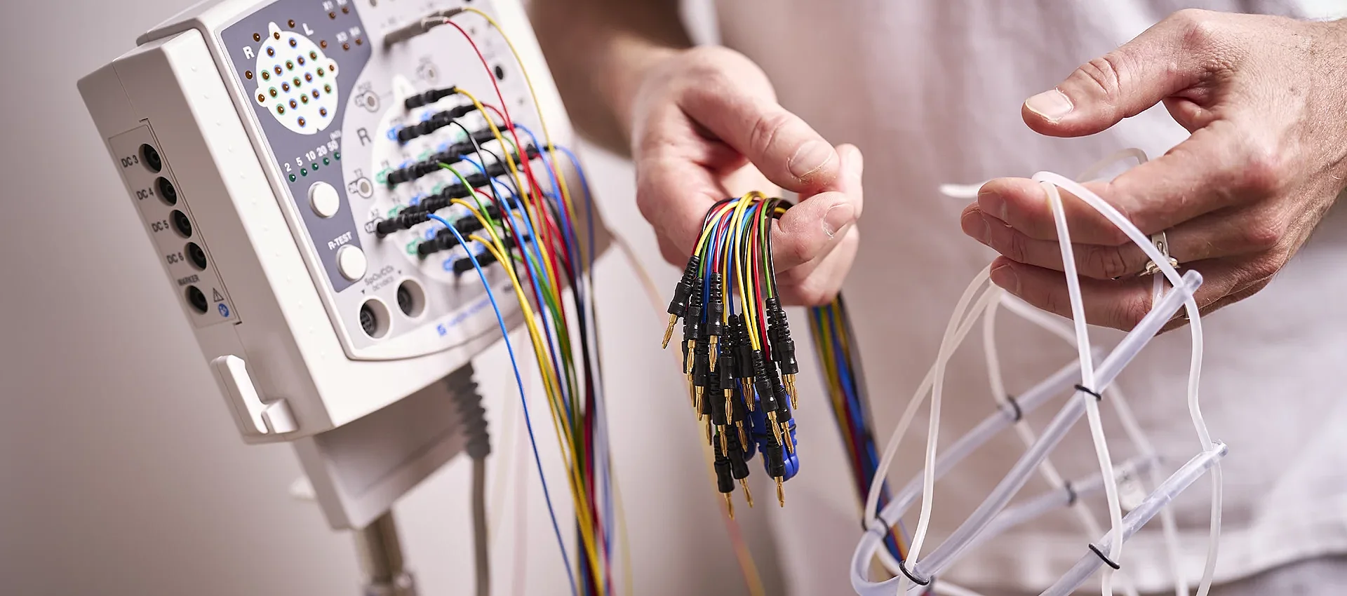
For these examinations, we use a latest-generation Nihon Kohden Neuropack X3 system.
Nerve and Muscle Ultrasound
Nerve and muscle ultrasound is a painless and radiation-free technique that provides high-resolution imaging of nerves and muscles. It complements electrophysiological testing and allows direct assessment of nerve entrapments, inflammation, injuries, tumors, and muscle disorders.
For these examinations, we use a Philips EPIQ Elite with a high-resolution mL26-8 ultrasound transducer. This specialized probe provides detailed imaging of even the smallest peripheral nerves and superficial muscles.
Electroencephalography (EEG)
Electroencephalography (EEG) is a well-established, painless and radiation-free method for measuring the electrical activity of the brain. It provides important information about brain function and is used particularly in the evaluation of epilepsy, seizures, and disorders of consciousness.
For these examinations, we use a latest-generation Nihon Kohden Neurofax EEG-1200 system. It provides high-quality digital EEG recordings and enables precise assessment of brain activity. We also use activation procedures such as hyperventilation (deep breathing) and photic stimulation (flashing lights).
Lumbar Puncture and Cerebrospinal Fluid Analysis
During a lumbar puncture, cerebrospinal fluid is collected from the lower back. Subsequent cerebrospinal fluid analysisprovides important information for the evaluation of disorders affecting the brain, spinal cord, and meninges.
It is particularly useful in the diagnosis of inflammatory and infectious diseases of the nervous system, autoimmune disorders such as multiple sclerosis, neurodegenerative diseases such as Alzheimer’s disease, and inflammation of blood vessels (vasculitis).
In certain conditions, lumbar puncture can also be used therapeutically to relieve increased pressure (therapeutic lumbar puncture). We perform the procedure on an outpatient basis in our practice, under sterile conditions and after a detailed discussion of the procedure.
Memory and Dementia Assessment
Memory and concentration problems should be evaluated by a neurologist at an early stage. The assessment includes a detailed medical history, a neurological examination, and standardized cognitive tests (e.g. MoCA, MMSE, and the Clock Drawing Test) to assess memory, attention, and other cognitive functions.
Additional investigations may include EEG, laboratory tests, cerebrospinal fluid analysis, brain imaging, and other diagnostic procedures. The aim is to identify treatable causes, diagnose dementia as early as possible, and enable individualized treatment.
Tilt Table Test
The tilt table test is a specialized examination used to evaluate dizziness, fainting (syncope), and disorders of cardiovascular regulation. During the test, the table is gradually tilted from a lying to an upright position while blood pressure, heart rate, and heart rhythm are continuously monitored.
The examination is performed under medical supervision and can help identify disorders of cardiovascular regulation and the autonomic nervous system as possible causes of fainting and unexplained loss of consciousness. Additional autonomic function testing may include heart rate variability (HRV) and sympathetic skin response (SSR).
Fundus and Optic Nerve Assessment
Neuro-ophthalmological assessment combines neurological and ophthalmological evaluation and plays an important role particularly in disorders of the optic nerve and visual pathways, as well as when increased intracranial pressure is suspected.
Examination of the back of the eye provides a direct view of the retina, optic disc, and blood vessels and can provide important information about neurological and vascular disorders. Using digital fundus photography, we obtain high-resolution images of these structures. For this purpose, we use an iCare DRSplus widefield fundus camera.
Ultrasound examination of the optic nerve and optic disc complements the assessment, particularly when increased intracranial pressure or optic nerve disorders are suspected. For this examination, we use a Philips EPIQ Elite with a high-resolution ultrasound transducer.